Anatomy explained: the bone your new teeth stand on
Full-mouth implant plans are drawn around a handful of structures: the sinus above the upper back teeth, the nerve inside the lower jaw, the cheekbone, and the bone that heals onto titanium. Knowing where they sit makes a treatment plan much easier to read.
Why anatomy decides the plan
Two people can both be told they need "full mouth implants" and end up with very different operations. The reason is rarely the teeth. It is the bone that is left after teeth are lost, and what lies next to it.
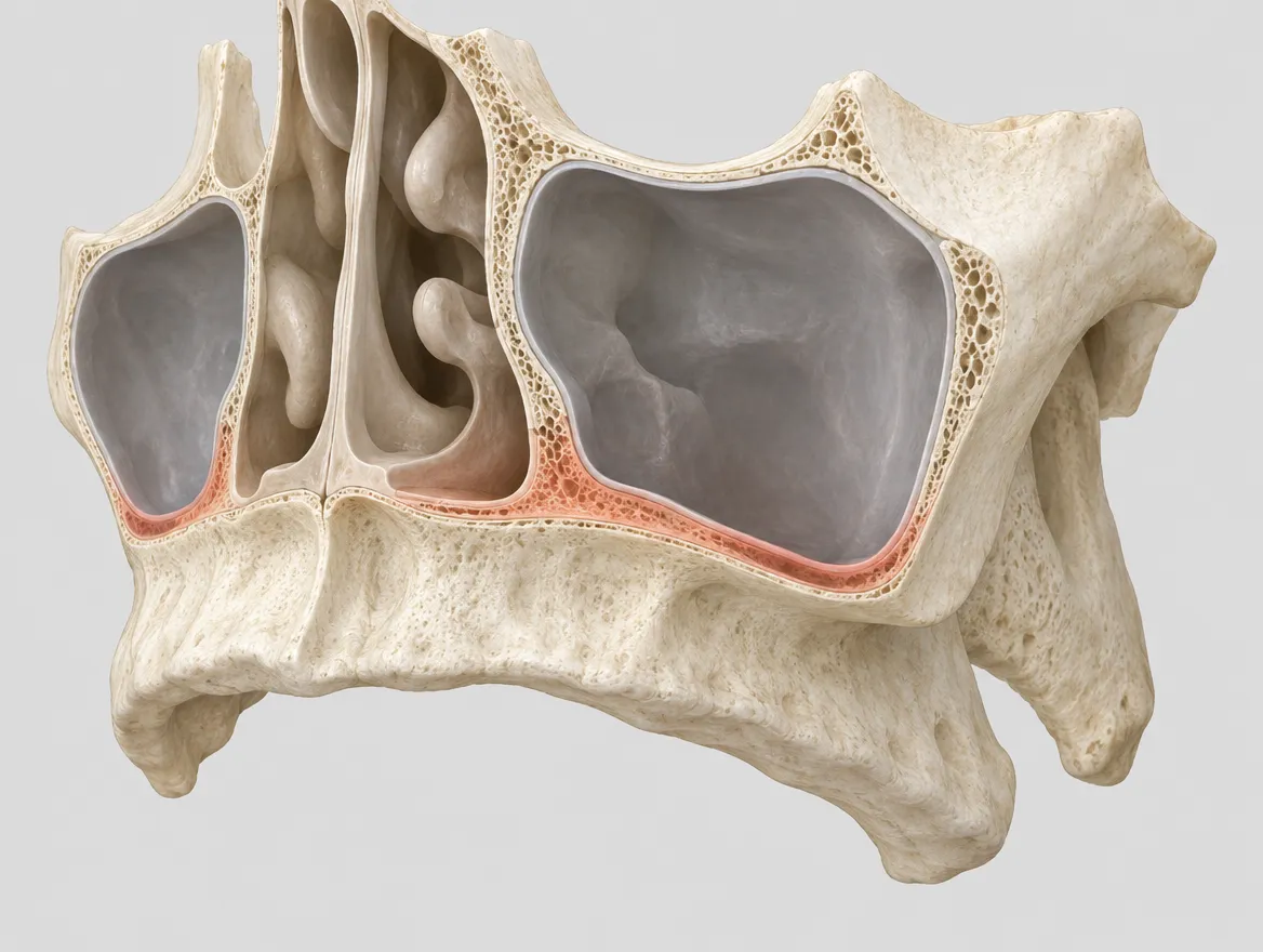
In the upper jaw the limit is usually the maxillary sinus, an air space that sits just above the roots of the back teeth and tends to grow downwards once those teeth are gone. In the lower jaw the limit is the inferior alveolar nerve, which runs through the bone and gives feeling to the lower lip and chin. When neither jaw has enough bone in the usual places, surgeons look further afield, to the cheekbone or the back of the upper jaw.
A 3D scan (CBCT) shows all of these in one picture. That is why Precious Dental plans every full-arch case on a 3D scan and tells you in writing how many implants are planned, which brand, and why.
The anatomy explainers
Lower jaw and nerve
The nerve canal, the mental foramen, the anterior loop and how often numbness happens.
Open →How implants fuse with bone
Primary and secondary stability, the week-three dip and why soft bone heals differently.
Open →Upper jaw and sinus
The sinus, soft upper bone, sinus lifts and the alternatives that avoid grafting.
Open →Cheekbone anchorage
Zygomatic implants: anatomy, surgical paths, survival and complication figures.
Open →The two jaws are not the same
Readers often assume an implant behaves the same anywhere in the mouth. The research says otherwise. The upper and lower jaws differ in bone density, in what lies next to them and in how they change after teeth are lost.
| Feature | Upper jaw (maxilla) | Lower jaw (mandible) | Read more |
|---|---|---|---|
| Main structure to avoid | Maxillary sinus above the back teeth | Inferior alveolar nerve and its exit at the mental foramen | Sinus · Nerve |
| Typical bone | Softer, more porous bone at the back | Denser bone, especially at the front | Healing |
| Ways to gain anchorage | Sinus lift, tilted implants, pterygoid or zygomatic implants | Tilted back implants in front of the nerve, short implants, grafting | Cheekbone |
| After severe bone loss | Ridge can become very thin and low | Ridge can flatten until the nerve lies near the surface | Bone-loss stages |
A large review of 94 publications found that implants placed in the softest category of bone failed more often than implants in any other bone type, and that missing bone volume was also linked to more failures (Chrcanovic et al., 2017). Soft bone is most often found in the back of the upper jaw, which is one reason surgeons often place more implants, or longer ones, in the upper arch.
Start with the jaw that worries you. If your dentist has mentioned a sinus lift or "not enough bone at the top", read the upper-jaw page first. If you have been warned about numbness, read the lower-jaw page. Unfamiliar words are in the glossary, and the studies behind the numbers are in the evidence library.
These explainers are general information, reviewed by our implant surgeon. They do not replace an examination: only your own scan shows where your sinus and nerve are.
Get your own full-mouth plan and price in writing
Send a recent panoramic X-ray or CBCT scan, or a few clear photos. Our implant surgeon reviews them and you receive an itemised plan in pounds, with approximate euro and dollar figures, before you book anything.