Jaw bone loss after tooth loss: the stages, and how fast it happens
When teeth go, the ridge of bone that held them starts to shrink. Dentists describe how far that has gone with a set of classes from 1988, and studies have measured how fast it happens.
 Medically reviewed by Dt. Hüseyin Yavuz Özbek, implant surgeon · Updated October 2026
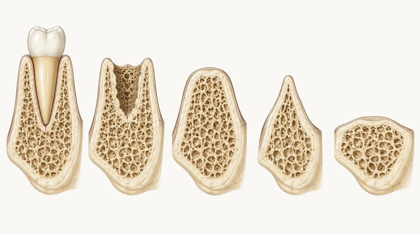
After a tooth is removed, the jaw ridge loses width faster than height: a review found horizontal loss of 29% to 63% and vertical loss of 11% to 22% within six months, with most of the change in the first three to six months. Over the longer term, ridges follow a predictable pattern described by the Cawood and Howell classes, from a well-rounded ridge to a thin knife-edge and finally a flat or sunken ridge. The later the stage, the more likely tilted implants, grafts or zygomatic implants are needed.
Why bone shrinks after tooth loss
The alveolar ridge exists to hold teeth. Once the roots are gone, the body starts to remodel it. The deep basal bone underneath stays fairly stable, but the ridge above it loses height and, especially, width Cawood, 1988. Dentists call this ridge resorption.
The first six months
A systematic review of 20 studies measured what happens to the ridge after an extraction Tan et al., 2012. Width went first.
Show the numbers as a table
| Item | Bone loss at 6 months | Source |
|---|---|---|
| Width (horizontal) | 3.79 mm | Tan et al., 2012 |
| Height, cheek side | 1.24 mm | Tan et al., 2012 |
| Height, front side (mesial) | 0.84 mm | Tan et al., 2012 |
| Height, back side (distal) | 0.8 mm | Tan et al., 2012 |
In percentage terms the same review reports 29% to 63% horizontal and 11% to 22% vertical loss at 6 months. Most change happens in the first 3 to 6 months.
In percentages, re-entry studies found horizontal bone loss of 29% to 63% and vertical loss of 11% to 22% six months after extraction. The reductions were rapid in the first three to six months and gradual after that. An earlier prospective study of back-tooth extractions also found that the major changes happen within the first year Schropp et al., 2003.
The thickness of the thin wall of bone in front of the roots matters. In front-tooth sites, walls of 1 mm or less lost about 7.5 mm of height, against 1.1 mm for thick walls Chappuis et al., 2017. A CBCT scan shows this wall before teeth are removed.
Most of these figures come from single-tooth extractions. When a whole arch of failing teeth is removed, the same biology applies across the jaw. This is one reason many full-arch plans place implants on the day of extraction, if the bone and the absence of active infection allow it.
The Cawood and Howell classes
In 1988, two surgeons studied 300 dried skulls and found that ridges change shape in a predictable sequence after tooth loss. They described it as a set of classes that dentists still use to talk about bone Cawood, 1988. The descriptions below follow the classes as they are commonly summarised from the original paper.
| Class | Ridge shape | What it often means for implants |
|---|---|---|
| I | Jaw with teeth | Teeth may still be saved; if not, implants can often go in at extraction |
| II | Straight after extraction (healing socket) | Bone is still close to its original volume; a window for implant placement |
| III | Well-rounded ridge, adequate height and width | Usually enough bone for standard implants |
| IV | Knife-edge ridge: adequate height, too narrow | Narrow implants, bone levelling, widening grafts or tilted implants |
| V | Flat ridge: too low and too narrow | Tilted or short implants, grafting, sinus lift in the upper jaw |
| VI | Depressed ridge with loss of basal bone | Major grafting; in the upper jaw, zygomatic implants may be considered |
The upper and lower jaws do not always match, and the front of a jaw can be a different class from the back. The upper back jaw has an extra limit, the maxillary sinus, which can expand downwards after tooth loss.
Options when bone is short
Thin or low bone does not automatically rule out fixed teeth. Common approaches include angling the back implants to reach better bone, using short implants, grafting, a sinus lift, or cheekbone anchorage. Where there is a little bone under the sinus, randomised trials found short implants (8 mm or less) had survival similar to longer implants in a lifted sinus (99.0% and 99.5% at 16 to 18 months), with about a third of the complications Thoma et al., 2015.
Bone around implants after treatment
Once implants have fused with the bone, studies show only small further changes. The first Swedish series reported an average of 1.5 mm during healing and the first year, then about 0.1 mm a year Adell et al., 1981.
Show the numbers as a table
| Item | Mean bone loss | Range | Source |
|---|---|---|---|
| Zirconia implants, 1 year | 0.79 mm | 0.73–0.86 mm | Pieralli et al., 2017 |
| Four implants, 3 years (review) | 1.3 mm | – | Patzelt et al., 2014 |
| Implants, 10+ years (review) | 1.3 mm | – | Moraschini et al., 2015 |
| Four implants, lower, 10 yrs | 1.72 mm | 1.59–1.85 mm | Maló et al., 2019 |
| Four implants, lower, 15 yrs | 2.32 mm | 1.98–2.66 mm | Maló et al., 2019 |
Measured from different reference points in different studies. The early Brånemark data reported 1.5 mm in healing and the first year, then about 0.1 mm a year.
Larger losses are usually a sign of peri-implantitis or overload rather than normal ageing, which is why baseline X-rays and regular checks matter. See complication rates.
What to ask about your own bone
- Has the plan been made from a 3D scan rather than a flat panoramic X-ray?
- Where exactly is bone short, and which technique is proposed for each area?
- If grafting or a sinus lift is suggested, how much extra healing time does it add, and what is the alternative? Our candidate checker lists the questions to bring to a consultation.
Common questions
Does bone keep shrinking for years after teeth are lost?
The fastest change is in the first three to six months after extraction, followed by slower, gradual reduction afterwards. That is why people who have worn dentures for decades often have much less bone than people whose teeth were removed recently.
Which Cawood and Howell class am I?
Only a 3D scan (CBCT) can tell, and the class can differ between the front and back of the same jaw, or between upper and lower jaws. Many full-arch patients have different classes in different areas.
Can lost bone be rebuilt?
Often, with grafting or a sinus lift, but these add healing time and cost. Alternatives include tilted implants that reach better bone, short implants and, in the upper jaw, zygomatic implants. Your surgeon chooses based on the scan.
Do implants stop bone loss?
Implants load the bone through chewing, and long-term studies show only small further changes around well-integrated implants, for example an average of 1.72 mm at 10 years in one four-implant study. They do not regrow bone that has already gone.
Why are implants often placed on the day teeth are removed?
Partly to use the bone while it is still there. Placing implants at the time of extraction, when the bone allows, avoids waiting months while the ridge narrows. Whether this is possible depends on infection, bone quality and how firm the implants are on the day.
Sources
- Cawood JI, Howell RA. A classification of the edentulous jaws. Int J Oral Maxillofac Surg 1988. PMID 3139793
- Tan WL, Wong TL, Wong MC, Lang NP. A systematic review of post-extractional alveolar hard and soft tissue dimensional changes in humans. Clin Oral Implants Res 2012. PMID 22211303
- Schropp L, Wenzel A, Kostopoulos L, Karring T. Bone healing and soft tissue contour changes following single-tooth extraction: a clinical and radiographic 12-month prospective study. Int J Periodontics Restorative Dent 2003. PMID 12956475
- Chappuis V, Araújo MG, Buser D. Clinical relevance of dimensional bone and soft tissue alterations post-extraction in esthetic sites. Periodontol 2000 2017. PMID 28000281
- Thoma DS, Zeltner M, Hüsler J, Hämmerle CH, Jung RE. EAO Supplement Working Group 4 - EAO CC 2015 Short implants versus sinus lifting with longer implants to restore the posterior maxilla: a systematic review. Clin Oral Implants Res 2015. PMID 25997901
- Adell R, Lekholm U, Rockler B, Brånemark PI. A 15-year study of osseointegrated implants in the treatment of the edentulous jaw. Int J Oral Surg 1981. PMID 6809663
- Maló P, de Araújo Nobre M, Lopes A, Ferro A, Botto J. The All-on-4 treatment concept for the rehabilitation of the completely edentulous mandible: A longitudinal study with 10 to 18 years of follow-up. Clin Implant Dent Relat Res 2019. PMID 30924309
- Patzelt SB, Bahat O, Reynolds MA, Strub JR. The all-on-four treatment concept: a systematic review. Clin Implant Dent Relat Res 2014. PMID 23560986
- Moraschini V, Poubel LA, Ferreira VF, Barboza Edos S. Evaluation of survival and success rates of dental implants reported in longitudinal studies with a follow-up period of at least 10 years: a systematic review. Int J Oral Maxillofac Surg 2015. PMID 25467739
- Pieralli S, Kohal RJ, Jung RE, Vach K, Spies BC. Clinical Outcomes of Zirconia Dental Implants: A Systematic Review. J Dent Res 2017. PMID 27625355
Checked October 2026. Where we give our own view as a clinic, we say so.
Get your own full-mouth plan and price in writing
Send a recent panoramic X-ray or CBCT scan, or a few clear photos. Our implant surgeon reviews them and you receive an itemised plan in pounds, with approximate euro and dollar figures, before you book anything.